Images of human clinical signs
Images on this page show the typical clinical signs of leptospirosis, namely conjunctival suffusion and a transient petechial rash. As the images we have created use CGI models there is no restriction of patient privacy, and so they may be used for any purpose, including publication online and in print, provided the visible copyright information and the embedded document information remains intact and unchanged.
The images below download in PNG format – click the thumbnails.
Petechial rash

This transient skin rash is seen in many (but not all) cases of leptospirosis and is identical to that seen in conditions such as bacterial meningitis. Red, irregular blotches appear on the skin that are dark red in color, sometimes turning a purple hue. They can appear anywhere on the body but in leptospirosis are often seen on the lower legs and the palatte. As with meninococcal rashes, pressure from a transparent object (such as a drinking glass) will blanch the sourrounding skin but the red blotches will remain unaltered.
The rash is caused by small blood vessels in the skin leaking blood into the tissues, where the blood forms a small red patch with irregular shape but quite sharp edges. As the color is from red blood cells that are unable to move, pressing on the skin does not change their color. The images above show a moderate to severe rash; in some patients only a small area of skin will be affected. The blotches will be in a range of sizes, up to 20mm diameter. In particularly severe infections the skin can break and cause some very slow bleeding from larger patches.
Conjuntival suffusion
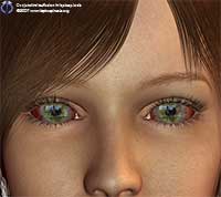
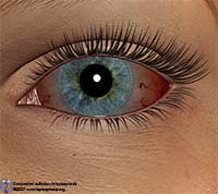
Conjunctical suffusion is a reddening of the conjunctiva (the front surface of the eye) caused by increased blood flow. Normally the conjunctiva’s blood supply is so small the vessels are invisible, but in leptospirosis these extremely small vessels become leaky, so red blood cells can enter the tissue of the conjunctiva. Flow is also increased in the vessels themselves as the immune response generates inflammation. It is visible against the sclera (the white of the eye) as both a general red tint and increased visibility of blood vessels. It is often painful and itchy, but unlike with primary conjuntivitis (commonly seen in allergies) there is little generation of excess tear fluid. In leptospirosis the effect will be similar in both eyes – where it displays only on one side it is more indicative of a local eye infection. Conjunctical suffusion is a very common sign and is expected in almost every patient to some extent.